Luke D. Lynch, Jinho Park, and Gheorghe Chistol (in press at Cell, 2026).
[Download PDF]
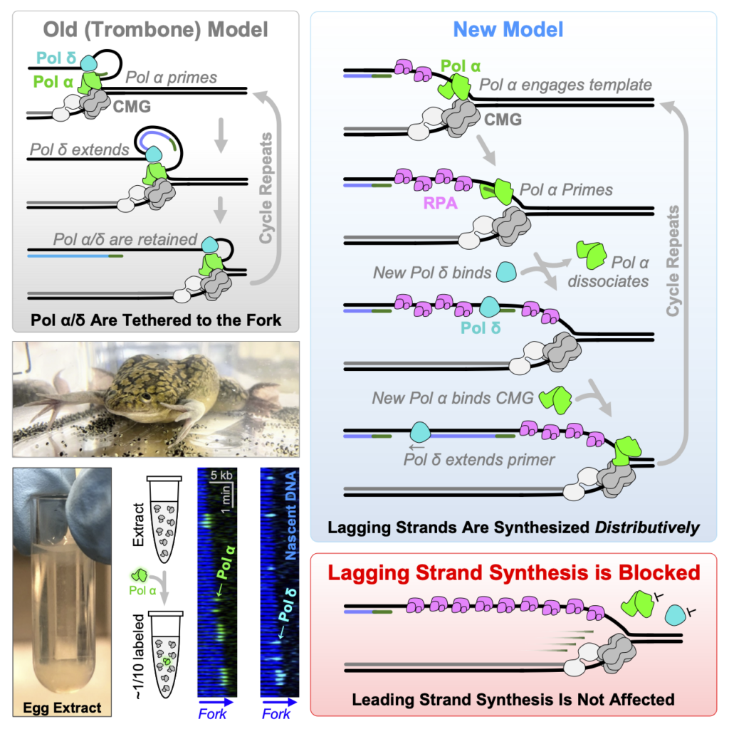
DNA’s anti-parallel structure presents a topological challenge to the replisome, as leading and lagging strands must be synthesized in opposite directions. How lagging strand synthesis is coordinated with leading strand synthesis remains unresolved in eukaryotes. To address this question, we directly visualized the dynamics of lagging strand polymerases Pol α and Pol δ at active replication forks using single-molecule imaging in Xenopus nuclear extracts. Surprisingly, we find that neither Pol α nor Pol δ is stably tethered to the replisome. Instead, lagging strand synthesis occurs distributively and entails the rapid recruitment of new molecules of Pol α and Pol δ for each Okazaki fragment. Consequently, leading and lagging strand synthesis are not intrinsically coupled. We propose that metazoans solve the problem of anti-parallel replication not by actively coordinating leading and lagging strand synthesis, but instead simply by recruiting new polymerases to synthesize each Okazaki fragment.
Riki Terui, Larissa Sambel, Jinho Park, Elyse Hsin-Lien Hwang, Benjamin Rhys Duewell, Dan Song, Gheorghe Chistol (in press at Nature Communications, 2026).
[Download PDF]
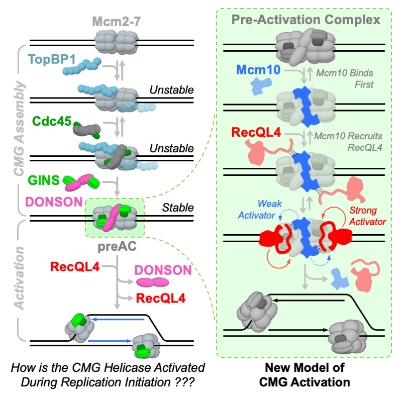
Chromosomes are copied from thousands of origins. At each origin, two replicative DNA helicases are first assembled, then activated to begin unwinding DNA. Several replication proteins are subsequently recruited to the active helicase, forming a replisome. The helicase must undergo dramatic conformational changes during its activation, and this process remains poorly understood, especially in metazoa. How the metazoan replicative helicase is activated, and which proteins promote this essential process are long-standing questions. Using a combination of single-molecule imaging and ensemble biochemistry, we show that Mcm10 and RecQL4 act in a concerted manner to activate replicative helicases. Mcm10 first binds to inactive helicases, then recruits RecQL4, and RecQL4 acts in tandem with Mcm10 to promote helicase activation. Like RecQL4, Mcm10 is not incorporated into replisomes and dissociates from origins during replication initiation. In the absence of Mcm10, RecQL4 is recruited to origins via an interaction with the Mcm7 subunit of the helicase. Our data reveal that Mcm10 and RecQL4 play partially redundant roles during helicase activation, help resolve long-standing controversies about the roles of Mcm10 and RecQL4 in DNA replication, and reveal replication initiation defects caused by pathologic RecQL4 mutations.
Riki Terui, Scott Berger, Larissa Sambel, Linda Song, and Gheorghe Chistol (Cell, 2024).
[Download PDF]
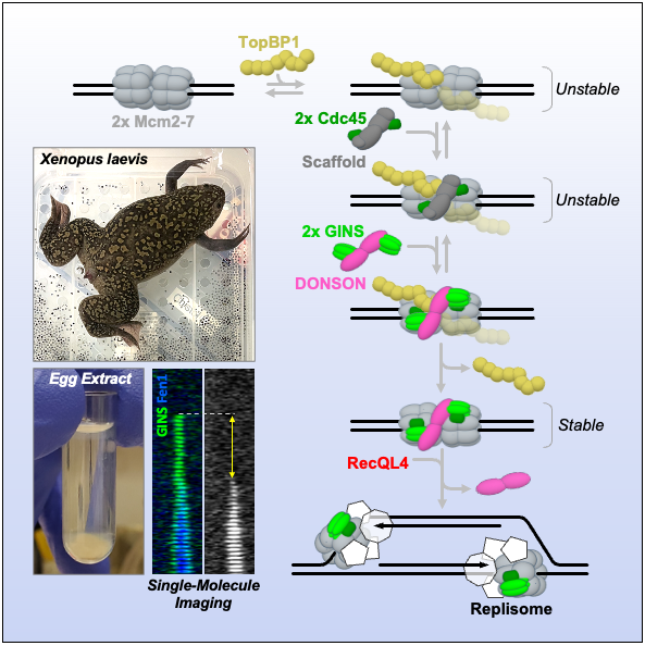
Metazoan genomes are copied bidirectionally from thousands of replication origins. Replication initiation entails the assembly and activation of two CMG helicases (Cdc45*Mcm2-7*GINS) at each origin. This requires several replication firing factors (including TopBP1, RecQL4, DONSON) whose exact roles are still under debate. How two helicases are correctly assembled and activated at each origin is a long-standing question. By visualizing the recruitment of GINS, Cdc45, TopBP1, RecQL4, and DONSON in real time, we uncovered that replication initiation is surprisingly dynamic. First, TopBP1 transiently binds to the origin and dissociates before the start of DNA synthesis. Second, two Cdc45 are recruited together, even though Cdc45 alone cannot dimerize. Next, two copies of DONSON and two GINS simultaneously arrive at the origin, completing the assembly of two CMG helicases. Finally, RecQL4 is recruited to the CMG*DONSON*DONSON*CMG complex and promotes DONSON dissociation and CMG activation via its ATPase activity.
Scott Berger and Gheorghe Chistol (Methods in Cell Biology, 2023).
[Download PDF]
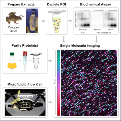
During cell division, the genome of each eukaryotic cell is copied by thousands of replisomes – large protein complexes consisting of several dozen proteins. Recent studies suggest that the eukaryotic replisome is much more dynamic than previously thought. To directly visualize replisome dynamics in a physiological context, we recently developed a single-molecule approach for imaging replication proteins in Xenopus egg extracts. These extracts contain all the soluble nuclear proteins and faithfully recapitulate DNA replication and repair in vitro, serving as a powerful platform for studying the mechanisms of genome maintenance. Here we present detailed protocols for conducting single-molecule experiments in nuclear egg extracts and preparing key reagents. This workflow can be easily adapted to visualize the dynamics and function of other proteins implicated in DNA replication and repair.
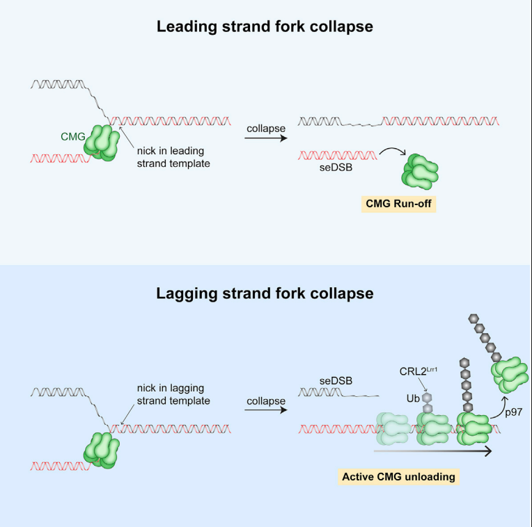
Vrtis K., Dewar J., Chistol G., Wu A.R., Graham T.G.W., Walter J.C. Molecular Cell, 81:1309-18 (2021). [PDF]
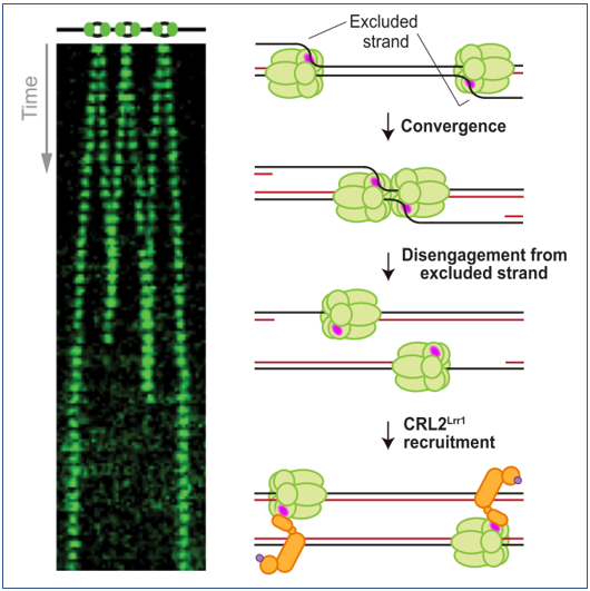
Low* E., Chistol* G [@]., Zaher M.S., Kochenova O.V., Walter J.C [@]. Genes & Development, 34:1534-45 (2020). [PDF]
* – these authors contributed equally.
[@] – corresponding authors
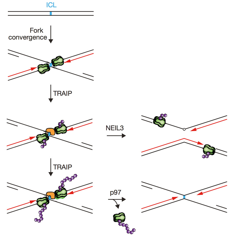
Wu R.A., Semlow D.R., Kamimae-Lanning A.N., Kochenova O.V., Chistol G. , Hodskinson M.R., Amunugama R., Sparks J.L., Wang M., Deng L., Mimoso C.A., Low E., Patel K.J., Walter J.C. Nature. 567(7747):267-72 (2019).
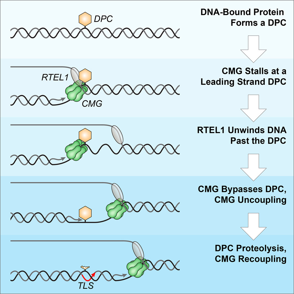
Sparks J.L.*, Chistol G.*, Gao A.O., Räschle M., Larsen N.B., Mann M., Duxin J.P., & Walter J.C. Cell, 176, 167-81.e21 (2019). [PDF]
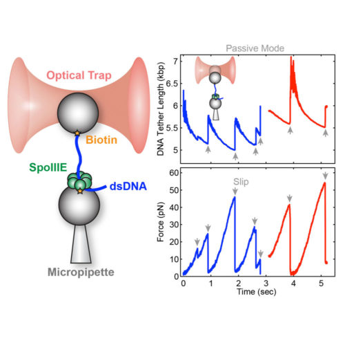
Liu N.*, Chistol G.*, Cui Y. & Bustamante C. eLife, 7 (2018). [PDF]
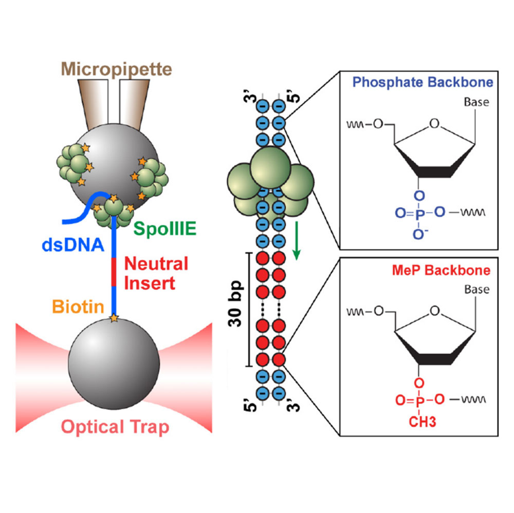
Liu N.*, Chistol G.* & Bustamante, C. eLife, 4 (2015). [PDF]
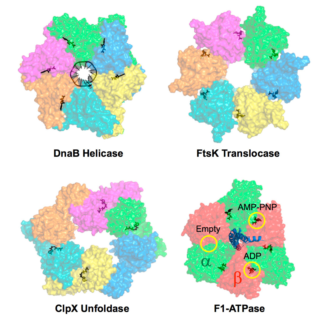
Liu S.*, Chistol G.* & Bustamante C. Biophysical Journal, 106, 1844–58 (2014). [PDF]
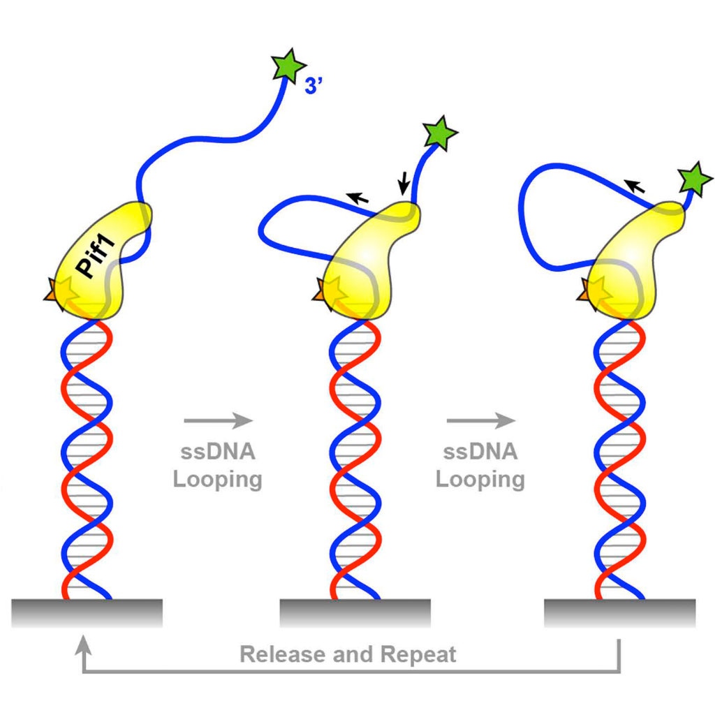
Chistol G. & Walter J.C. eLife 3, e02854 (2014). [PDF]
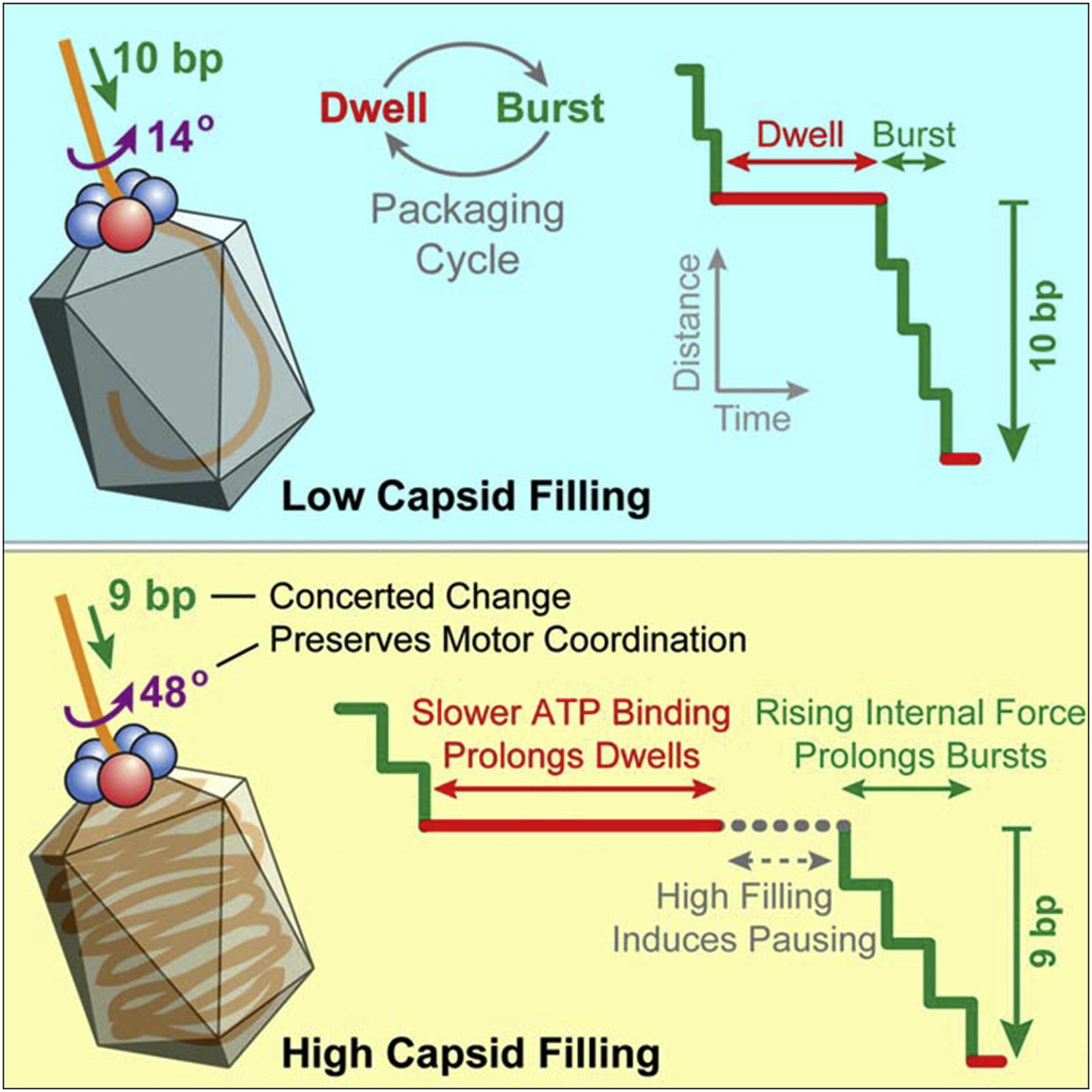
Liu S.*, Chistol G.*, Hetherington C.L.*, Tafoya S., Aathavan K., Schnitzbauer J., Grimes S., Jardine P.J., & Bustamante C. Cell 157, 702–13 (2014). [PDF]
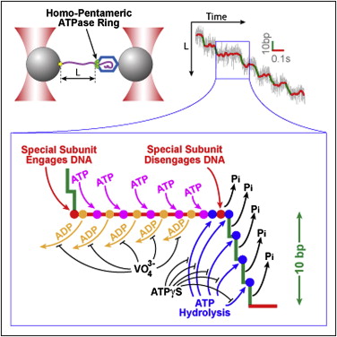
Chistol G.*, Liu S.*, Hetherington C.L., Moffitt J.R., Grimes S., Jardine P.J., & Bustamante C. Cell 151, 1017–28 (2012). [PDF]
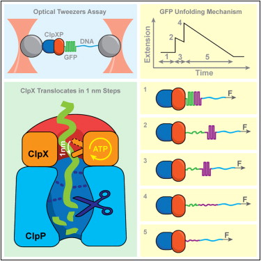
Maillard R.A., Chistol G., Sen M., Righini M., Tan J., Kaiser C.M., Hodges C., Martin A., & Bustamante C. Cell 145, 459–69 (2011). [PDF]
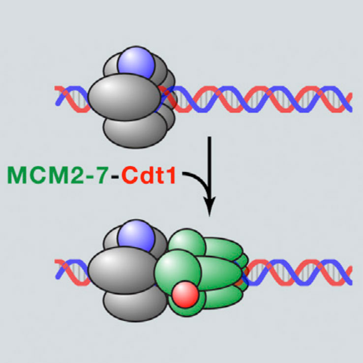
Chistol G. & Walter J.C. Cell 161, 429-30 (2015). [PDF]
